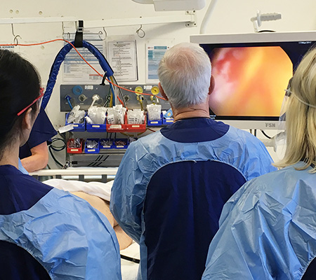
If you are diagnosed with bowel cancer, you will firstly need to have tests to determine the size of the cancer tumour, its position and whether it has spread. This process is known as 'staging'.
Your specialist needs to know the extent (stage) of the disease to plan the best treatment.
Sometimes however staging is not complete until after surgery to remove the tumour.
The stage is based on whether the tumour has invaded nearby tissues, whether the cancer has spread and, if so, to what parts of the body.
Your specialist may order some of the following tests to find out the cancer's stage, so staging may not be complete until all of the tests have been completed:
Blood tests: Your specialist checks for carcinoembryonic antigen (CEA) and other substances in the blood. Some people who have bowel cancer or other conditions have a high CEA level. You may also have blood test that measures chemicals that are normally found in your liver, known as a Liver Function Test. An abnormal result can be a sign the cancer has spread to the liver.


One of the tools specialists use to describe the stage is the TNM system.
TNM is a shorthand description of the staging classification, which helps specialists to understand very quickly what your cancer looks like.
The results from your diagnostic tests and scans are used to answer the following questions:
Colonoscopy: If colonoscopy was not performed for diagnosis, your specialist checks for abnormal areas along the entire length of the colon and rectum with a colonoscope.
Endorectal ultrasound: An ultrasound probe is inserted into your rectum. The probe sends out sound waves that people cannot hear. The waves bounce off your rectum and nearby tissues, and a computer uses the echoes to create a picture. The picture may show how deep a rectal tumour has grown or whether the cancer has spread to lymph nodes or other nearby tissues.
Chest x-ray: X-rays of your chest may show whether cancer has spread to your lungs.
CT scan: An x-ray machine linked to a computer takes a series of detailed pictures of areas inside your body. You may receive an injection of dye. A CT scan may show whether cancer has spread to the liver, lungs, or other organs.
PET-CT scan: A CT scan can be combined with a PET scan which is a medical imaging technique which produces a three-dimensional, colour image of your body. When taken together, the results can be combined to show where there are any cell changes in the body, and whether the cancer has spread.
MRI scan: Magnetic resonance imaging (MRI) uses magnetic and radio waves (not X-rays) to show the tumour(s) in great detail and look at the blood supply to the liver. During the scan you will have to lie in the scanner for up to an hour, and, whilst it is very noisy, it is painless. Let the doctors know in advance if you are claustrophobic. You may be asked to drink a liquid 'contrast medium' before a CT or MRI scan, or be given an injection of a contrast medium during the scan (which may give you a hot flush for a few minutes). The dye travels to your liver to help produce a better image.


One of the tools specialists use to describe the stage is the TNM system.
TNM is a shorthand description of the staging classification, which helps specialists to understand very quickly what your cancer looks like.
The results from your diagnostic tests and scans are used to answer the following questions:
- Tumour (T): Has the tumour grown into the wall or the colon or rectum? How many layers?
- Node (N): Has the tumour spread to the lymph nodes? If so, where and how many?
- Metastasis (M): Has the cancer metastasized to other parts of the body? If so, where and how much?
The results are combined to determine the stage of your cancer.
There are five (5) stages: stage 0 (zero) and stages I through IV (1 through to 4).
You may also hear about the Dukes' system or the Australian Clinico-Pathological Staging (ACPS) system.
Ask your specialist to explain the stage of your cancer in a way you can understand. This will help you to choose the best treatment for you.


Tumour (T)
Using the TNM system, the "T" plus a letter or number (0 to 4) is used to describe how deeply the primary tumour has grown into the bowel lining.
Some stages are also divided into smaller groups that help describe the tumour in even more detail.
- TX: The primary tumour cannot be evaluated.
- T0: There is no evidence of cancer in the colon or rectum.
- Tis: Refers to carcinoma in situ (also called cancer in situ). Cancer cells are found only in the epithelium or lamina propria, which are the top layers lining the inside of the colon or rectum.
- T1: The tumour has grown into the submucosa, which is the layer of tissue underneath the mucosa or lining of the colon.
- T2: The tumour has grown into the muscularis propria, a deeper, thick layer of muscle that contracts to force along the contents of the intestines.
- T3: The tumour has grown through the muscularis propria and into the subserosa, which is a thin layer of connective tissue beneath the outer layer of some parts of the large intestine, or it has grown into tissues surrounding the colon or rectum.
- T4a: The tumour has grown into the surface of the visceral peritoneum, which means it has grown through all layers of the colon.
- T4b: The tumour has grown into or has attached to other organs or structures.

Node (N)
The 'N' in the TNM system stands for lymph nodes.
The lymph nodes are tiny, bean-shaped organs located throughout the body.
Lymph nodes help the body fight infections as part of the immune system.
Lymph nodes near the colon and rectum are called regional lymph nodes. All others are distant lymph nodes that are found in other parts of the body.
- NX: The regional lymph nodes cannot be evaluated.
- N0: There is no spread to regional lymph nodes.
- N1a: There are tumour cells found in 1 regional lymph node.
- N1b: There are tumour cells found in 2 to 3 regional lymph nodes.
- N1c: There are nodules made up of tumour cells found in the structures near the colon that do not appear to be lymph nodes.
- N2a: There are tumour cells found in 4 to 6 regional lymph nodes.
- N2b: There are tumour cells found in 7 or more regional lymph nodes.
Metastasis (M)
The 'M' in the TNM system describes cancer that has spread to other parts of the body, such as the liver or lungs. This is called distant metastasis.
- MX: Distant metastasis cannot be evaluated.
- M0: The disease has not spread to a distant part of the body.
- M1a: The cancer has spread to 1 other part of the body beyond the colon or rectum, but not to distant parts of the peritoneum (the lining of the abdominal cavity).
- M1b: The cancer has spread to more than 1 part of the body other than the colon or rectum, but not to distant parts of the peritoneum.
-
M1c: The cancer has spread to distant parts of the peritoneum, and may or may not have spread to another part of the body beyond the colon or rectum.
Stage 0 (Carcinoma in Situ) | Tis, N0, M0
In stage 0, abnormal cells are found in the mucosa (innermost layer) of the bowel wall.
These abnormal cells may become cancer and spread. Stage 0 is also called carcinoma in situ.


In Stage I, cancer has formed in the mucosa (innermost layer) of the bowel wall and has spread to the submucosa (T1), and it may also have grown into the muscularis propria (T2).
Cancer may have spread to the muscle layer of the bowel wall.
It has not spread into nearby tissue or lymph nodes (N0) or to distant sites (M0)


Stage II bowel cancer is divided into stage IIA, stage IIB, and stage IIC.
- Stage IIA: Cancer has spread through the muscle layer of the bowel wall to the serosa (outermost layer) of the bowel wall but has not gone through them (T3). It has not reached nearby organs and has not spread to nearby lymph nodes (N0) or to distant sites (M0).
- Stage IIB: Cancer has spread through the serosa (outermost layer) of the bowel wall but has not grown into other nearby tissues or organs (T4a). It has not yet spread to nearby lymph nodes (N0) or to distant sites (M0).
-
Stage IIC: Cancer has spread through the serosa (outermost layer) of the bowel wall and is attached to or grown into other nearby tissues or organs (T4b). It has not yet spread to nearby lymph nodes (N0) or to distant sites (M0).

Stage III bowel cancer is divided into stage IIIA, stage IIIB, and stage IIIC.
- Stage IIIA: Cancer has grown through the mucosa into the submucosa (T1), and it may also have grown into the muscularis propria (T2). It has spread to 1 to 3 nearby lymph nodes (N1) or into areas of fat near the lymph nodes but not the nodes themselves (N1c). It has not spread to distant sites (M0).
OR
The cancer has grown through the mucosa into the submucosa (T1). It has spread to 4 to 6 nearby lymph nodes (N2a). It has not spread to distant sites (M0).

- Stage IIIB: The cancer has grown into the outermost layers of the bowel (T3) or through the viscerarl peritoneum (T4a), but has not reached nearby organs. It has spread to 1 to 3 nearby lymph nodes (N1a or N1b) or into areas of fat near the lymph nodes but not the nodes themselves (N1c). It has not spread to distant sites (M0).
OR
The cancer has grown into the muscularis propria (T2) or into the outermost layers of the bowel (T3). It has spread to 4 to 6 nearby lymph nodes (N2a). It has not spread to distant sites (M0).
OR
The cancer has grown through the mucosa into the submucosa (T1), and it may also have grown into the muscularis propria (T2). It has spread to 7 or more nearby lymph nodes (N2b). It has not spread to distant sites (M0).

- Stage IIIC: The cancer has grown through the wall of the bowel (including the visceral peritoneum) but has not reached nearby organs (T4a). It has spread to 4 to 6 nearby lymph nodes (N2a). It has not spread to distant sites (M0).
OR
The cancer has grown into the outmost layers of the bowel wall (T3) or through the visceral peritoneum (T4a) but has not reached nearby organs. It has spread to 7 or more nearby lymph nodes (N2b). It has not spread to distant sites (M0).
OR
The cancer has grown through the bowel wall and is attached to or has grown into other nearby tissues or organs (T4b). It has spread to at least one nearby lymph node or into areas of fat near the lymph nodes (N1 or N2). It has not spread to distant sites (M0).

Stage IV (also known as metastatic, advanced or secondary) colon cancer is divided into stage IVA, stage IVB and stage IVC.
- Stage IVA: The cancer may or may not have grown through the bowel wall (any T). It might or might not have spread to nearby lymph nodes (any N). It has spread to 1 distant organ (such as the liver or lung) or distant set of lymph nodes, but not to distant parts of the peritoneum (the lining of the abdominal cavity) (M1a).
- Stage IVB: The cancer might or might not have grown through the bowel wall (any T). It migh or might not have spread to nearby lymph nodes (any N). It has spread to more than 1 distant organ (such as the liver or the lung) or distant set of lymph nodes, but not to distant parts of the peritoneum (M1b).
-
Stage IVC: The cancer might or might not have grown through the bowel wall (any T). It might or might not have spread to nearby lymph nodes (any N). It has spread to distant parts of the peritoneum, and may or may not have spread to distant orgas or lymph nodes (M1c).

Recurrent bowel cancer is cancer that has recurred (come back) after it has been treated.
The cancer may come back in the bowel or in other parts of the body, such as the liver, lungs, or both.
If the cancer does return, there will be another round of tests to learn about the extent of the recurrence.
Source: National Cancer Institute


